Book Appointment Now
Understanding Bullous Diseases: Navigating Blistering Skin Conditions
The skin is our body’s largest organ and its primary defense against the environment. When the delicate cohesion between the layers of the skin is disrupted, the result is often the formation of blisters—known medically as “bullae.” While a small blister from a tight shoe is a common, harmless occurrence, bullous diseases represent a group of serious, often chronic, and sometimes life-threatening conditions where the body’s own immune system attacks the very structures that hold the skin together.
These diseases are complex, rare, and often debilitating. Understanding them requires looking beneath the surface—literally—to the molecular bonds that maintain the integrity of our skin and mucous membranes.
What Are Bullous Diseases?
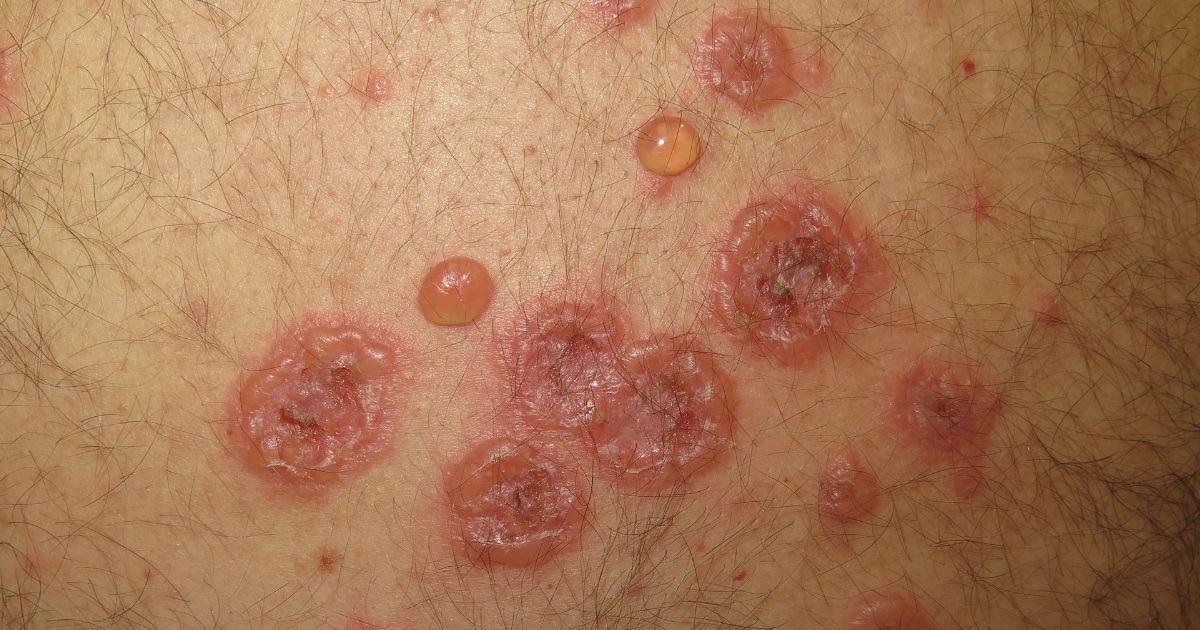
Bullous diseases are characterized by the formation of blisters (bullae) that are greater than 0.5 cm in diameter. These conditions are generally divided into two main categories: autoimmune bullous diseases and inherited (genodermatosis) bullous diseases.
In autoimmune bullous diseases, which are the most clinically significant in dermatology, the body produces autoantibodies that target specific proteins (desmosomes or hemidesmosomes) responsible for skin adhesion. This process is called acantholysis (loss of cell-to-cell adhesion) or subepidermal separation, resulting in the top layer of skin lifting away from the layers beneath it, filling with fluid, and creating a blister.
Key Autoimmune Bullous Conditions
1. Pemphigus Vulgaris (PV)
Pemphigus vulgaris is perhaps the most well-known and severe of the autoimmune blistering diseases. It is characterized by thin-walled, flaccid blisters that rupture easily, leaving behind painful, raw erosions.
- Target: The immune system attacks desmogleins, the proteins that act as “glue” between skin cells (keratinocytes).
- Impact: It often begins in the mouth, sometimes months before skin lesions appear, making eating and speaking extremely painful. If left untreated, the surface area of the erosions can become life-threatening due to infection and fluid loss.
2. Bullous Pemphigoid (BP)
Unlike PV, bullous pemphigoid is more common in the elderly. The blisters here are tense, thick-walled, and do not rupture as easily as those in pemphigus.
- Target: The immune system attacks the hemidesmosomes, which anchor the epidermis to the basement membrane (the layer beneath the skin).
- Impact: It typically presents with intense itching (pruritus), which may precede the appearance of actual blisters by weeks or months. While still serious, it is generally easier to manage than pemphigus vulgaris.
3. Mucous Membrane Pemphigoid (Cicatricial Pemphigoid)
This variant primarily affects the mucous membranes—the eyes, nose, throat, and genitals. The danger here is not just the blistering, but the subsequent scarring (cicatrization). In the eyes, for instance, this can lead to symblepharon (adhesion of the eyelid to the eyeball), which can cause permanent vision loss if not addressed aggressively.
4. Dermatitis Herpetiformis (DH)
Dermatitis herpetiformis is a unique, intensely itchy skin manifestation of Celiac disease. It appears as clusters of small, fluid-filled bumps, often on the elbows, knees, and buttocks. It is caused by the body’s reaction to dietary gluten, which triggers the formation of IgA immune complexes that deposit in the skin.
The Mechanism: Why Do Blisters Form?
To understand bullous diseases, one must visualize the skin structure. The epidermis (outer layer) is anchored to the dermis (inner layer) by a basement membrane zone. Within the epidermis, cells are locked together by protein bridges.
- Intraepidermal (Pemphigus group): When the “glue” between cells is destroyed, the cells pull apart, creating a space within the epidermis. This causes the soft, fragile blisters seen in Pemphigus Vulgaris.
- Subepidermal (Pemphigoid group): When the “anchor” holding the epidermis to the dermis is destroyed, the entire top layer lifts off. This creates a stronger, “tense” blister, as the entire skin thickness is separating from the underlying dermis.
Diagnosis: The “Gold Standard”
Diagnosing a bullous disease requires more than just a visual examination. Because many of these diseases look similar, dermatologists rely on a three-pronged diagnostic approach:
- Clinical Presentation: Evaluating the location, type of blister, and involvement of mucous membranes.
- Skin Biopsy (Histopathology): A small sample of skin is taken to see where the separation is occurring (within the skin or at the base).
- Direct Immunofluorescence (DIF): This is the gold standard. A biopsy sample is stained with special antibodies that glow under a microscope, allowing the doctor to see the exact pattern of immune system attack (the “fingerprint” of the disease).
Management and Treatment Strategies
Historically, bullous diseases were often fatal. Today, the development of immunosuppressive therapies has turned these into manageable, chronic conditions.
- Systemic Corticosteroids: The traditional “first-line” treatment. High doses of prednisone are used to quickly stop the immune attack, though long-term use carries significant side effects.
- Steroid-Sparing Agents: Medications like azathioprine, mycophenolate mofetil, or methotrexate are often introduced to allow doctors to lower the dose of steroids over time.
- Rituximab: A biological therapy that has revolutionized the treatment of Pemphigus. It targets the B-cells responsible for producing the harmful autoantibodies, often leading to long-term remission.
- Dietary Management: For patients with Dermatitis Herpetiformis, a strict gluten-free diet is the most effective treatment, often eliminating the need for medication entirely.
Living with a Bullous Disease
Living with a bullous disease is an exercise in vigilance. Patients must be careful to avoid skin trauma, as even minor friction can cause new blisters to form. Furthermore, because these patients are often on immunosuppressive therapy, they are at higher risk for secondary infections, making hygiene and regular monitoring vital.
The emotional toll should not be overlooked. The pain associated with mucosal lesions, the distress of constant skin erosions, and the side effects of long-term medication can significantly impact quality of life. Peer support groups and counseling are essential components of the care plan.
Conclusion
Bullous diseases remind us of the incredible precision required by our immune system to maintain our health. When that precision fails, the results can be devastating. However, thanks to advancements in immunology and targeted biological therapies, the prognosis for patients with these conditions has improved dramatically.
If you or a loved one experience unexplained, persistent blistering or raw, painful sores in the mouth or on the skin, it is critical to seek the expertise of a dermatologist. Early diagnosis is the most powerful tool in preventing extensive skin damage and ensuring a better long-term outcome.
Disclaimer: This article is for educational purposes only and does not constitute medical advice, diagnosis, or treatment. If you are experiencing symptoms, please consult a medical professional immediately.

Dr. S. N. Gupta is a renowned skin specialist and dermatologist in Prayagraj, with extensive experience in diagnosing and treating various skin, hair, nail and Sexually Transmitted Diseases conditions for last 28 years. Address Shree Balaji Medical Traffic Chauraha, Rajapur Prayagraj Mob 9415613743