Book Appointment Now
Porokeratosis : Types, Symptoms, Causes, and Management – Dr S.N. Gupta
When unusual, ring-shaped skin lesions with distinct, raised borders begin to appear on your skin, it can be a cause for concern. One rare group of conditions that presents this way is known medically as porokeratosis.
While the name may sound unfamiliar, understanding porokeratosis is essential because certain variants carry a recognized risk of developing into secondary skin cancers if left unmonitored.
This comprehensive guide explores everything you need to know about porokeratosis, including its various clinical types, root causes, symptoms, diagnosis, and modern management strategies.
What Is Porokeratosis?
Porokeratosis is a rare, chronic disorder of keratinization—the process by which skin cells mature and move to the surface. It is characterized by abnormal clones of epidermal cells that expand outward, forming distinct patches or plaques surrounded by a signature elevated ridge.
The Hallmark Feature: The Cornoid Lamella
The defining microscopic feature that unites all forms of porokeratosis is the cornoid lamella. Under a dermatological microscope, this appears as a thin, tightly packed column of parakeratotic cells (cells that retain their nuclei longer than normal) rising through the outer layer of the skin (stratum corneum). Clinically, this translates into the sharp, raised ridge or “wall” that rings the outer edge of every porokeratosis lesion.
The Different Types of Porokeratosis
Porokeratosis is not a single disease, but rather a family of distinct clinical variants categorized by how and where the lesions appear on the body:
1. Porokeratosis of Mibelli (PM)
- Appearance: Usually begins in childhood or early adolescence as small, brownish papules that slowly expand over years into larger plaques with a well-defined, raised keratosic border.
- Common Locations: Typically appears on the extremities, hands, feet, and occasionally the face or mucous membranes.
2. Disseminated Superficial Actinic Porokeratosis (DSAP)
- Prevalence: This is the most common form of porokeratosis.
- Triggers: Strongly driven by ultraviolet (UV) sun exposure. Lesions typically erupt during early adulthood or middle age after years of cumulative sun exposure.
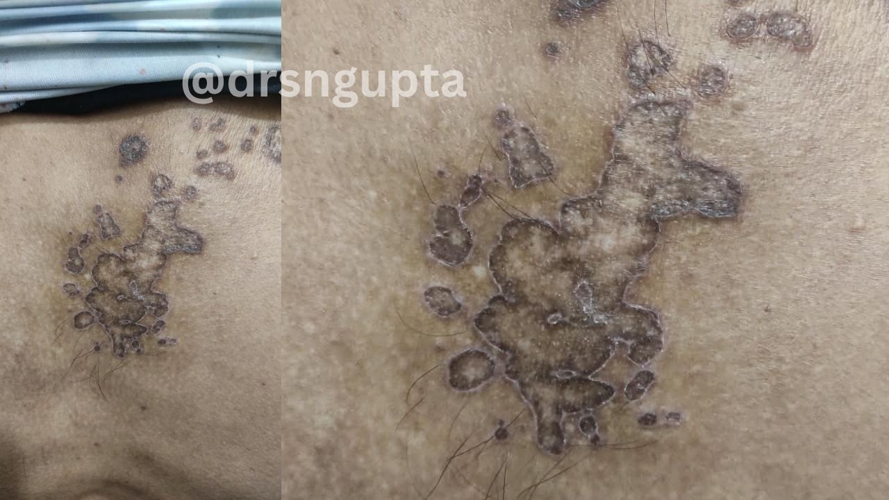
- Appearance: Numerous small, annular (ring-shaped), pink-to-brown flat spots with a delicate raised rim.
- Common Locations: Sun-exposed areas such as the lower legs, arms, and shoulders, usually sparing the face, palms, and soles.
3. Disseminated Superficial Porokeratosis (DSP)
- Similar to DSAP, but unlike its actinic counterpart, DSP is not strictly linked to sun exposure. Lesions can appear across both sun-exposed and sun-protected areas of the body (such as the trunk and abdomen).
4. Porokeratosis Palmaris et Plantaris Disseminata (PPPD)
- Appearance: Features tiny, punctate (pinpoint) pits or rings that specifically target the palms of the hands and the soles of the feet before spreading outward to the limbs and trunk. It often begins in childhood or adolescence.
5. Linear Porokeratosis
- Appearance: The rarest variant, characterized by linear streaks or whorls of lesions that follow the natural embryonic developmental pathways of the skin (known as Blaschko’s lines). It typically presents at birth or in early childhood.
What Causes Porokeratosis?
The exact underlying cellular mechanism involves a genetic mutation that causes certain epidermal keratinocytes to proliferate abnormally and cornify improperly.
Key contributing factors include:
1. Genetic Mutations and Familial Links
Many forms of porokeratosis—particularly autosomal dominant variants—are inherited. Mutations in genes related to the mevalonate pathway (such as MVD or PMVK) are frequently implicated, disrupting normal cell cholesterol synthesis and keratinocyte differentiation.
2. Ultraviolet (UV) Radiation
For DSAP and sun-aggravated forms of porokeratosis, UV light acts as a major environmental trigger. Sun exposure damages cellular DNA, activating abnormal keratinocyte clones that manifest as characteristic ring lesions on sun-soaked skin.
3. Immunosuppression
Individuals with compromised immune systems—such as organ transplant recipients, people living with HIV, or those undergoing long-term immunosuppressive drug therapies—are at a significantly higher risk of developing widespread or aggressive forms of porokeratosis.
Symptoms and What Porokeratosis Looks Like
Porokeratosis lesions have a very distinct visual presentation that helps dermatologists make an initial clinical assessment:
- Ring-Shaped (Annular) Plaques: Lesions typically feature a central area that may look normal, slightly atrophic (thinned), or brownish, enclosed by a hard, raised, ridged border (the cornoid lamella).
- Asymptomatic or Mildly Itchy: Most lesions are physically painless, though some patients experience mild itching, dryness, or a burning sensation, especially when exposed to heat or friction.
- Slow Expansion: Individual spots tend to slowly expand outward over time while maintaining their central cleared or scaly zone.
The Cancer Risk: Why Monitoring Matters
One of the most critical aspects of porokeratosis is its classification as a precancerous condition.
While many lesions remain stable and benign for a lifetime, all forms of porokeratosis carry a documented risk of malignant transformation. Over time, chronic cellular instability within the plaques can progress into skin cancers, most commonly squamous cell carcinoma (SCC), and occasionally basal cell carcinoma or melanoma.
Red Flag Signs of Malignancy:
- Rapid, aggressive enlargement of a lesion.
- A spot that begins bleeding spontaneously, ulcerates, or develops a hard, nodular center.
- Persistent pain or tenderness in a previously painless plaque.
Because of this risk, individuals diagnosed with porokeratosis—especially DSAP or long-standing Mibelli types—require regular, lifelong skin examinations by a board-certified dermatologist.
Professional Diagnosis and Medical Evaluation
Diagnosing porokeratosis requires clinical expertise because its ring-like patterns can sometimes mimic other skin conditions like ringworm (tinea corporis), eczema, or psoriasis.
1. Clinical Examination with Dermoscopy
A dermatologist will examine the skin using a specialized magnifying tool called a dermatoscope. Under polarized light, the distinct, bright white or hyperpigmented peripheral track corresponding to the cornoid lamella is often clearly visible.
2. Skin Biopsy
To confirm an ambiguous diagnosis or rule out early malignancy, the doctor may perform a small punch biopsy. Examining the tissue under a microscope allows the pathologist to identify the pathognomonic cornoid lamella within the epidermis.
Treatment and Management Options
Because porokeratosis is a chronic, deep-seated genetic condition, curing it completely can be challenging. Treatment goals focus on reducing symptoms, improving cosmetic appearance, and aggressively monitoring or destroying suspicious lesions to prevent skin cancer.
1. Topical Therapies
- Topical Retinoids: Prescription creams (like tretinoin or tazarotene) help normalize skin cell turnover and flatten the raised borders.
- 5-Fluorouracil (5-FU) or Imiquimod: These topical chemotherapeutic or immune-response modifier creams are frequently prescribed for widespread DSAP to destroy abnormal dysplastic cells.
- Topical Vitamin D Analogs: Calcipotriene is sometimes used to regulate keratinocyte growth.
2. Destructive Procedures (For Isolated Lesions)
- Cryotherapy: Freezing individual lesions with liquid nitrogen can destroy abnormal superficial tissue.
- Curettage and Desiccation: Scraping away the lesion combined with mild electrocautery.
- Laser Therapy: Ablative lasers (such as CO2 or Erbium:YAG lasers) are effective at vaporizing stubborn or thick plaques, particularly in porokeratosis of Mibelli.
3. Rigorous Sun Protection (Crucial for DSAP)
Because UV radiation acts as a primary catalyst and mutational driver for DSAP:
- Apply a broad-spectrum sunscreen with an SPF of 30 or higher daily, rain or shine.
- Wear sun-protective clothing, wide-brimmed hats, and seek shade during peak UV hours (10 AM to 4 PM).
Can You Treat Porokeratosis at Home?
Never attempt to treat, scrape, or burn porokeratosis lesions at home.
Attempting DIY removal with over-the-counter wart removers, sharp blades, or harsh chemical oils can lead to:
- Severe chemical burns and secondary bacterial infections.
- Permanent, unsightly scarring or dyspigmentation.
- Delayed Cancer Detection: Damaging or masking a lesion at home can obscure early malignant changes, allowing a developing squamous cell carcinoma to go unnoticed until it becomes advanced.
When to See a Dermatologist
Schedule an appointment with a qualified dermatologist promptly if you notice:
- New, ring-shaped, scaly spots spreading across your arms, legs, or trunk.
- An existing porokeratosis lesion that changes color, thickens, bleeds, or develops an open sore that refuses to heal.
- A family history of skin cancer combined with persistent, sun-induced skin spots.
Frequently Asked Questions (FAQs)
Is porokeratosis contagious?
No. Porokeratosis is an inherited or acquired disorder of keratinization and cellular turnover. It is not caused by a virus, fungus, or bacteria, and you cannot spread it to other people or catch it from someone else.
Will porokeratosis go away on its own?
Rarely. Porokeratosis is typically a chronic, lifelong skin condition. While individual lesions may occasionally stabilize, they do not spontaneously disappear without medical intervention.
How serious is the cancer risk?
While malignant transformation occurs in a minority of cases, studies estimate that roughly 7% to 10% of patients with certain forms of porokeratosis may develop squamous cell carcinoma within a lesion over their lifetime. Regular dermatological surveillance minimizes this risk significantly.
The Takeaway
Porokeratosis is a complex group of rare skin disorders marked by unique cornoid lamellae and ring-shaped lesions. While many forms are benign and manageable, their potential to progress into skin cancer makes professional medical supervision vital. If you notice unusual ringed patches on your skin, skip the home remedies and partner with a board-certified dermatologist for an accurate diagnosis, a personalized treatment plan, and routine skin cancer screenings.

Dr. S. N. Gupta is a renowned skin specialist and dermatologist in Prayagraj, with extensive experience in diagnosing and treating various skin, hair, nail and Sexually Transmitted Diseases conditions for last 28 years. Address Shree Balaji Medical Traffic Chauraha, Rajapur Prayagraj Mob 9415613743