Book Appointment Now
Acrodermatitis Enteropathica: Pathogenesis, Clinical Manifestations, Diagnosis, and Management
Introduction and Historical Perspective
Acrodermatitis enteropathica (AE) is a rare, severe, autosomal recessive metabolic disorder characterized by a primary defect in intestinal zinc absorption.Left untreated, the condition historically proved fatal, particularly during infancy.
The clinical entity was first described independently by Brandt in 1936 and later characterized in detail by Norwegian dermatologists Niels Danbolt and Karen Closs in 1942 and 1943. For decades, the etiology remained elusive, and patients were subjected to various ineffective regimens, including frequent hospitalizations, empirical antibiotics, and derivatives like diiodohydroxyquinoline (iodoquinol), which offered unpredictable and occasionally toxic symptomatic relief.
A monumental breakthrough occurred in 1973 when E.J. Moynahan linked the clinical manifestations of AE to profound zinc deficiency and successfully treated a dying infant with oral zinc supplementation.This discovery instantly transformed a progressive, lethal pediatric syndrome into a fully manageable condition with a near-perfect prognosis, provided lifelong therapy is maintained.
The molecular mechanism was completely elucidated in 2002, when researchers mapped the genetic locus to chromosome 8q24 and identified mutations in the SLC39A4 gene, which encodes the ZIP4 zinc transporter.
Etiology and Genetics
Acrodermatitis enteropathica can manifest in either an inborn (congenital) form or an acquired form. While the phenotypic expressions of both forms are nearly identical, their underlying root causes differ fundamentally.
1. Congenital Acrodermatitis Enteropathica
The congenital variant is inherited in an autosomal recessive pattern, meaning an affected individual inherits one mutated allele from each carrier parent.The disease stems from loss-of-function mutations in the SLC39A4 gene located on chromosome 8q24.
The SLC39A4 gene encodes the ZIP4 protein (Zrt- and Irt-like protein 4), a critical transmembrane transporter protein heavily expressed on the apical brush border of enterocytes within the duodenum and upper jejunum. ZIP4 is responsible for the active uptake of dietary zinc from the intestinal lumen. When ZIP4 is structurally or functionally compromised due to missense, nonsense, frameshift, or splice-site mutations, active zinc transport fails.
Consequently, even though an affected infant may consume adequate amounts of zinc, the body cannot absorb it, triggering systemic zinc deprivation.
2. Acquired Acrodermatitis Enteropathica
Acquired zinc deficiency syndrome can mimic congenital AE in every clinical detail. It occurs when normal zinc transporters are overwhelmed or bypassed due to external factors:
- Dietary Inadequacy:Exclusively breastfed infants born to mothers with low zinc content in their breast milk (due to transient neonatal zinc deficiency or maternal genetic mutations affecting zinc excretion into milk) can develop acquired AE.
- Malabsorptive States:Conditions that impair intestinal absorption, such as Crohn’s disease, celiac disease, bariatric surgery, or extensive small bowel resection, can precipitate severe zinc deficiency.
- Iatrogenic Depletion:Patients on total parenteral nutrition (TPN) whose regimens lack adequate supplemental zinc rapidly develop acquired AE-like symptoms within weeks.
- Increased Physiological Demand: States of high cell turnover, catabolism, burns, or chronic renal disease with excessive urinary loss can also drop systemic zinc reserves below critical thresholds.
Pathophysiology of Zinc Deficiency
Zinc ($Zn^{2+}$) is an essential trace element required for fundamental cellular operations. It acts as a structural or catalytic cofactor for more than 300 metalloenzymes and transcription factors. It is vital for DNA replication, RNA transcription, cell division, protein synthesis, and immune system modulation.
When zinc homeostasis collapses, widespread multi-organ dysfunction ensues:
- Epithelial and Epidermal Integrity: Zinc is indispensable for rapid keratinocyte proliferation and differentiation. Deficiency causes premature keratinization, parakeratosis, and structural breakdown of epidermal barriers, manifesting as characteristic skin lesions.
- Immunological Deficits: Zinc deficiency impairs both cell-mediated and humoral immunity. It causes thymic atrophy, decreases natural killer cell activity, suppresses neutrophil chemotaxis, and reduces lymphocyte proliferation, leaving patients highly vulnerable to bacterial (especially Staphylococcus aureus) and fungal (especially Candida albicans) superinfections.
- Gastrointestinal Dysfunction: The rapidly dividing enterocytes of the gastrointestinal tract suffer structural atrophy, leading to flattened mucosal villi, impaired digestive enzyme production, malabsorption, and chronic diarrhea.
- Neurosensory Alterations: Zinc plays an essential role in central nervous system development and neurotransmission. Depletion leads to irritability, emotional lability, depression, and sensory deficits like hypogeusia (reduced taste) and hyposmia (reduced smell).
Clinical Manifestations
The classic presentation of congenital acrodermatitis enteropathica features a diagnostic triad: acral and periorificial dermatitis, alopecia, and diarrhea.However, fewer than 20% of patients present with all three features simultaneously at onset.
1. Timing of Onset
In congenital AE, symptoms typically do not appear immediately at birth because neonates are protected by maternal zinc stores accumulated during the third trimester, coupled with the high bioavailability of zinc in breast milk. Symptoms characteristically emerge during weaning, when breast milk is replaced by cow’s milk or formula. Cow’s milk contains casein complexes and phytates that bind zinc tightly, reducing its bioavailability. In formula-fed infants, symptoms may manifest even earlier.
2. Cutaneous Findings (Dermatitis)
The skin eruption is the hallmark of the condition and typically evolves through distinct morphological stages:
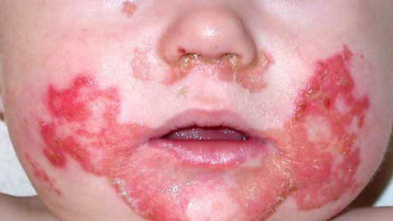
- Distribution:Lesions appear in symmetrically distributed, sharply demarcated, erythematous, eczematous, or psoriasiform plaques.They heavily target periorificial areas (around the mouth, eyes, nose, and perineal/anogenital regions) and acral regions (hands, feet, elbows, knees, ankles, and buttocks).
- Morphology:The initial presentation involves dry, scaly patches that rapidly progress to vesiculobullous, pustular, erosive, and crusted plaques. Secondary candidal or bacterial colonization is frequent, giving lesions a weeping, foul-smelling appearance.
- Nail Involvement:Paronychia (inflammation of the nail folds) and nail dystrophy (ridged, brittle, or completely shed nails) frequently accompany the acral rash.
3. Gastrointestinal Manifestations
Chronic, watery diarrhea is an early and persistent feature.It can range from mild loose stools to severe steatorrhea (fatty, foul-smelling stools) accompanied by abdominal distension, anorexia, and severe malabsorption, further compounding systemic malnutrition and growth failure.
4. Hair Changes (Alopecia)
Diffuse or patchy alopecia affecting the scalp, eyebrows, and eyelashes is common.The hair shafts become dry, brittle, lusterless, and easily pull out.
5. Systemic and Neuropsychiatric Symptoms
- Behavioral Changes:Affected infants are frequently described as profoundly irritable, apathetic, inconsolably cranky, and lethargic.
- Growth Retardation: Prolonged zinc deficiency halts linear growth and delays bone maturation, leading to severe failure to thrive in infants and children.
- Ophthalmologic Signs:Photophobia, blepharitis, conjunctivitis, and corneal opacities may develop.
- Immunological Vulnerability: Recurrent opportunistic infections of the skin, respiratory, and gastrointestinal tracts are major sources of morbidity and mortality. In rare instances, cutaneous viral infections like Kaposi’s varicelliform eruption can complicate the clinical picture.
Differential Diagnosis
Because its presentation mimics several other pediatric and dermatological conditions, diagnosing acrodermatitis enteropathica requires careful clinical differentiation from:
- Atopic Dermatitis / Eczema: While common, eczema rarely presents with the sharp periorificial and acral demarcation, nor is it typically accompanied by severe chronic diarrhea and alopecia.
- Candidiasis / Diaper Dermatitis: Fungal diaper dermatitis affects the diaper area, but true AE extends past the folds into perianal, perineal, and acral zones alongside systemic signs of malabsorption.
- Cystic Fibrosis: Can present with failure to thrive, diarrhea, and secondary essential fatty acid or zinc deficiencies due to pancreatic exocrine insufficiency.
- Biotinidase Deficiency (Multiple Carboxylase Deficiency): Shares features of periorificial dermatitis and alopecia, but is distinguished by neurological abnormalities (seizures, hypotonia) and organic aciduria.
- Essential Fatty Acid Deficiency: Results in scaly dermatitis and growth failure, usually seen in patients on fat-free parenteral nutrition.
Diagnostic Evaluation
Establishing a definitive diagnosis relies on a combination of clinical suspicion, laboratory assays, and genetic testing.
Clinical Suspicion (Periorificial/Acral Rash, Diarrhea, Alopecia)
        │
        ├──> Laboratory Testing: Serum/Plasma Zinc Levels (Low: <50 µg/dL)
        ├──> Biochemical Marker: Alkaline Phosphatase (Low levels support diagnosis)
        └──> Confirmatory Testing: SLC39A4 Gene Sequencing (Identifies ZIP4 mutations)
1. Laboratory Biomarkers
- Serum/Plasma Zinc Levels: Total plasma or serum zinc levels are markedly depressed. Normal reference ranges generally span 70 to 130 µg/dL (10.7 to 19.9 µmol/L). In active AE, levels frequently drop below 50 µg/dL.Caveat: Serum zinc constitutes less than 0.1% of total body zinc and can be falsely lowered by hypoalbuminemia, acute-phase systemic inflammation, or stress. Furthermore, improper sample collection using non-acid-washed containers can lead to environmental zinc contamination and false normal readings.
- Alkaline Phosphatase (ALP):Serum alkaline phosphatase is a zinc-dependent metalloenzyme.Depressed ALP activity serves as a reliable, highly sensitive surrogate marker for functional zinc deficiency, often remaining low even when serum zinc levels are borderline or inconclusive.
2. Histopathology
A skin biopsy of an active lesion is non-specific but helpful for ruling out other pathologies. Histological examination typically reveals:
- Confluent parakeratosis.
- Psoriasiform epidermal hyperplasia.
- Extensive intracellular edema and pallor within the upper spinous layer (pallor of keratinocytes).
- Necrotic keratinocytes and subcorneal or intraepidermal pustules containing neutrophils.
3. Genetic Testing
Molecular genetic testing via sequence analysis of the SLC39A4 gene on chromosome 8q24 provides definitive confirmation of congenital acrodermatitis enteropathica.Identifying compound heterozygous or homozygous pathogenic variants confirms the diagnosis, eliminates diagnostic ambiguity, and aids in genetic counseling for families.
Management and Treatment
The treatment of acrodermatitis enteropathica is remarkably straightforward, highly effective, and life-saving.
1. Zinc Supplementation
- Formulation:Oral elemental zinc is the therapy of choice. Various salts are available, including zinc sulfate, zinc gluconate, and zinc acetate. Zinc sulfate is the most widely utilized and cost-effective formulation.(Note: 4 mg of zinc sulfate contains approximately 1 mg of elemental zinc; clinicians must always calculate dosage based strictly on elemental zinc content).
- Dosing:The standard therapeutic dosage for both congenital and acquired AE is 3 mg/kg/day of elemental zinc for infants and children, divided into two or three daily doses to maximize gastrointestinal tolerance and absorption. For adults with acquired forms, dosages range from 50 to 150 mg of elemental zinc daily.
- Duration: In congenital AE caused by SLC39A4 mutations, lifelong zinc supplementation is mandatory. Stopping therapy inevitably leads to a rapid relapse of symptoms within weeks. For acquired forms, supplementation is continued until the underlying malabsorptive trigger is resolved or dietary intake is optimized.
2. Clinical Response Timeline
The therapeutic response to oral zinc is often dramatic:
- Within 24 to 72 hours: Irritability, lethargy, and mood disturbances rapidly resolve.
- Within 1 to 2 weeks:Diarrhea ceases, appetite normalizes, and skin erosions begin re-epithelializing at the edges.
- Within 4 to 8 weeks: Complete resolution of cutaneous plaques, normalization of hair growth (alopecia reversal), and restoration of normal nail plates.
3. Adjunctive Care
During acute presentations, topical therapy may be employed alongside systemic zinc:
- Gentle barrier creams, emollients, and soothing wet compresses assist with re-epithelialization.
- Short courses of targeted topical or systemic antimicrobials/antifungals are indicated if secondary bacterial (Staphylococcus) or fungal (Candida) superinfections complicate open skin erosions.
Monitoring and Potential Complications
While zinc supplementation is transformative, ongoing medical supervision is vital to avoid complications related to chronic high-dose therapy or unmonitored trace element imbalances.
1. Copper Deficiency (Hypocupremia)
Zinc and copper share common intestinal absorption pathways utilizing divalent metal transporters (such as metallothionein induction in enterocytes). High-dose, long-term oral zinc supplementation induces intestinal metallothionein synthesis, which binds copper preferentially and blocks its absorption. This can precipitate secondary copper deficiency, leading to microcytic anemia, leukopenia, neutropenia, and severe neurological deterioration. To prevent this, baseline and periodic monitoring of serum copper and ceruloplasmin levels is essential.
2. Routine Laboratory Surveillance
Patients on lifelong zinc therapy require regular check-ups, typically every 6 to 12 months, involving:
- Serum zinc and alkaline phosphatase levels.
- Complete blood counts (CBC) to monitor for copper-deficiency anemia or leukopenia.
- Serum copper and lipid profiles.
- Evaluation for gastrointestinal tolerance (as high doses of zinc salts can cause transient nausea, vomiting, or abdominal cramping).
Prognosis
Before the discovery of zinc therapy, acrodermatitis enteropathica was a progressive, debilitating disease that frequently resulted in death during early childhood due to severe secondary infections and systemic wasting.
In modern clinical practice, the prognosis is excellent.With prompt diagnosis and strict adherence to lifelong oral zinc supplementation, patients experience complete clinical remission, normal physical and cognitive development, and a normal life expectancy. Women with congenital AE who become pregnant may experience increased metabolic demands and temporary exacerbations, requiring carefully monitored upward adjustments of their zinc dosage during gestation to protect both maternal health and fetal development.
Conclusion
Acrodermatitis enteropathica stands as a textbook medical paradigm illustrating how identifying a single biochemical deficiency can revolutionize patient outcomes. By recognizing the classic triad of periorificial/acral dermatitis, diarrhea, and alopecia—and confirming the diagnosis via plasma zinc levels, alkaline phosphatase screening, and SLC39A4 genetic testing—clinicians can intervene decisively.Lifelong oral zinc supplementation completely reverses the devastating systemic pathology of the disease, ensuring that affected individuals lead healthy, normal lives.

Dr. S. N. Gupta is a renowned skin specialist and dermatologist in Prayagraj, with extensive experience in diagnosing and treating various skin, hair, nail and Sexually Transmitted Diseases conditions for last 28 years. Address Shree Balaji Medical Traffic Chauraha, Rajapur Prayagraj Mob 9415613743