
Book Appointment Now
Lipoid Proteinosis: A Comprehensive Medical Review – Dr S. N. Gupta
Introduction
Lipoid Proteinosis (LP), also known as Urbach-Wiethe disease or hyalinosis cutis et mucosae, is a rare autosomal recessive genetic disorder characterized by the widespread deposition of hyaline material in the dermis and mucous membranes. First systematically described by the dermatologist Erich Urbach and the otolaryngologist Camillo Wiethe in 1929, this condition manifests through a diverse array of dermatological, otolaryngological, and neurological symptoms.
Although fewer than 400 cases have been documented in medical literature worldwide, Lipoid Proteinosis remains a subject of significant clinical interest due to its distinct phenotypic presentation, predictable early onset, and the diagnostic challenges it presents to clinicians. This article provides an in-depth examination of the molecular etiology, pathophysiology, clinical manifestations, diagnostic strategies, and current management paradigms associated with lipoid proteinosis.
Etiology and Molecular Genetics
Lipoid Proteinosis is caused by loss-of-function mutations in the Extracellular Matrix Protein 1 (ECM1) gene, located on the long arm of chromosome 1q21.3.
The Role of ECM1
The ECM1 gene encodes a glycoprotein that plays a critical role in:
- Extracellular matrix homeostasis: Interacting with structural components such as collagen types I and III, perlecan, and fibronectin.
- Angiogenesis: Regulating endothelial cell proliferation and vascular stability.
- Dermal-epidermal adhesion: Supporting the structural integrity of the basement membrane zone.
- Keratinocyte differentiation: Modulating epidermal growth and maturation.
Mutations in ECM1—which include missense, nonsense, frameshift, and splice-site alterations—lead to a deficiency or functional impairment of the ECM1 protein. Without adequate ECM1 to regulate basement membrane turnover and extracellular matrix deposition, aberrant hyaline-like material accumulates progressively within the papillary and reticular dermis, as well as around blood vessels and adnexal structures. This accumulation results in the characteristic thickening and hardening of affected tissues.
Inheritance Pattern
LP follows an autosomal recessive inheritance pattern. Affected individuals inherit two mutated copies of the ECM1 gene, one from each carrier parent. Consanguinity is a frequently noted epidemiological factor in families with LP, increasing the probability of homozygous mutations. Heterozygous carriers are typically asymptomatic but carry a 25% risk per pregnancy of producing an affected offspring.
Pathophysiology and Histopathology
The hallmark pathological feature of lipoid proteinosis is the extracellular deposition of periodic acid-Schiff (PAS)-positive, diastase-resistant hyaline material.
Microscopic Findings
- Skin Biopsy: Histological examination reveals marked thickening of the papillary dermis and the walls of dermal blood vessels. Hyaline deposits are characteristically found surrounding blood vessels, eccrine sweat glands, and hair follicles.
- Electron Microscopy: Demonstrates reduplication of the basement membrane zone and deposition of amorphous, finely fibrillar electron-dense material.
- Biochemical Composition: The deposited material consists primarily of non-collagenous glycoproteins, lipids, and various basement membrane components (such as laminin and type IV collagen), reflecting the impaired matrix turnover caused by ECM1 deficiency.
Clinical Manifestations
The clinical presentation of lipoid proteinosis is remarkably variable, though symptoms typically manifest in early infancy and progress through childhood before stabilizing in adulthood. The organ systems most commonly involved include the skin, oral cavity, upper respiratory tract, and central nervous system.
+-------------------------------------------------------------------------+
|                  CLINICAL SPECTRUM OF LIPOID PROTEINOSIS                |
+-------------------------------------------------------------------------+
|  1. Dermatological: Moniliform blepharitis, acneiform scars, warty skin |
|  2. Otolaryngological: Hoarseness (first sign), macroglossia, stridor   |
|  3. Neurological: Temporal lobe calcifications, seizures, psychiatric   |
+-------------------------------------------------------------------------+
1. Dermatological Manifestations
Skin changes are often among the earliest and most recognizable signs of the disease:
- Hoarse Cry in Infancy: Often the very first presenting symptom, caused by hyaline infiltration of the true vocal cords shortly after birth.
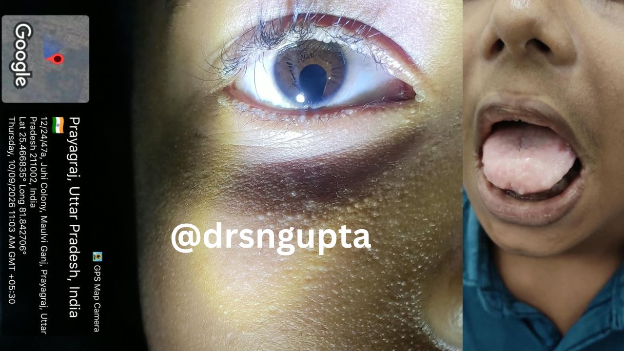
- Blistering and Scarring: Recurrent vesiculobullous eruptions occur on trauma-prone areas (such as the face, hands, and elbows) during childhood, healing with varioliform (pox-like) or acneiform scars.
- Infiltration and Thickening: The skin gradually develops a yellowish-white, waxy, or thickened appearance. Skin laxity is frequently lost, giving the face a prematurely aged look (elastosis serpiginosa-like changes).
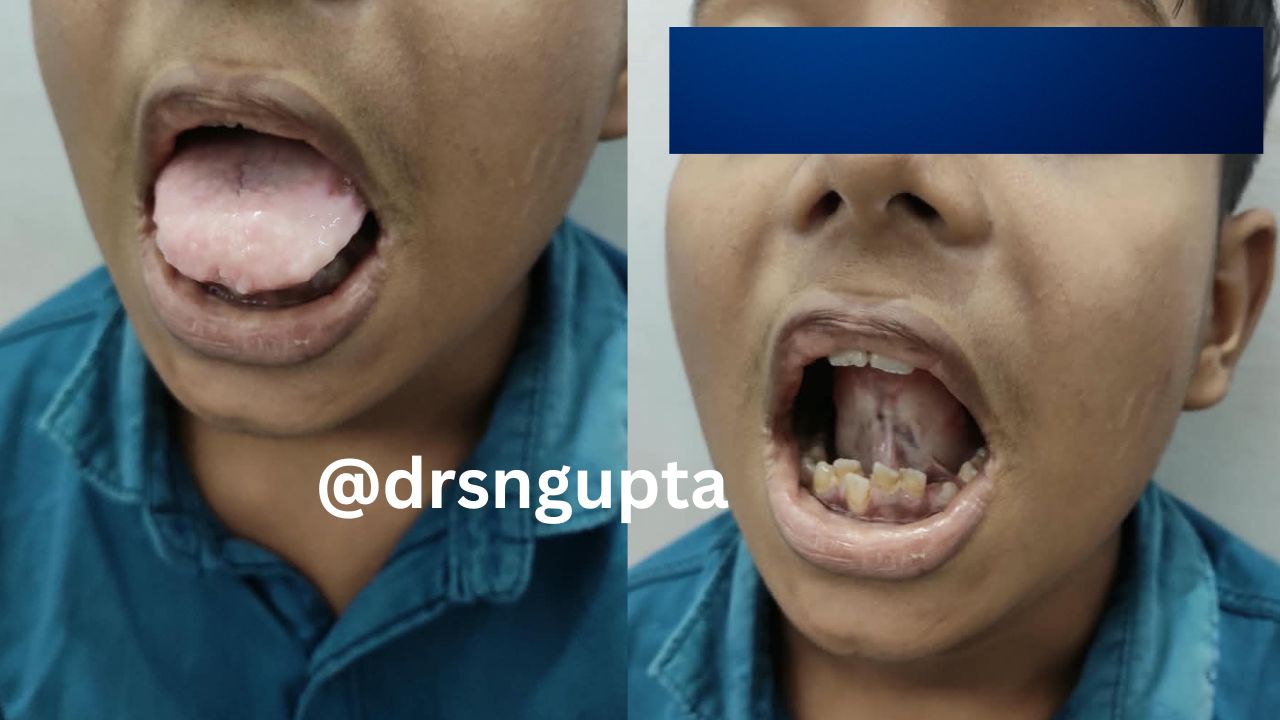
- Moniliform Blepharitis: A pathognomonic feature consisting of a string of small, bead-like papules arrayed along the free margins of the eyelids.
- Alopecia: Diffuse or patchy hair loss can occur due to follicular involvement by hyaline deposits.
2. Otolaryngological Manifestations
- Voice Alteration: Persistent hoarseness progresses to severe dysphonia or absolute aphonia due to stiffening, thickening, and nodular infiltration of the vocal cords.
- Oral Cavity Involvement: Infiltration of the tongue (macroglossia) and frenulum leads to restricted tongue mobility, difficulty in protrusion, and subsequent speech impediments. The mucosa of the soft palate, tonsils, and pharynx becomes stiff, thick, and yellowish-white.
- Dental Abnormalities: Enamel hypoplasia, delayed eruption, and oligodontia (congenital absence of certain teeth) are occasionally reported.
- Airway Compromise: Severe infiltration of the upper respiratory tract can occasionally precipitate life-threatening laryngeal obstruction, necessitating emergency tracheostomy.
3. Neurological and Psychiatric Manifestations
Neurological involvement occurs in approximately 20% to 50% of patients and is typically linked to intracranial calcifications:
- Intracranial Calcifications: Symmetrical, comma-shaped or bean-shaped calcifications within the bilateral amygdala and medial temporal lobes are classic radiographic findings.
- Seizure Disorders: Temporal lobe epilepsy can develop secondary to amygdala calcification and structural irritation.
- Neuropsychiatric Disturbances: Anxiety, depression, emotional lability, memory deficits, and schizophrenia-like psychosis have been documented, correlating with structural limbic system pathology.
Differential Diagnosis
Because lipoid proteinosis shares clinical features with several other dermatological and metabolic disorders, a careful differential diagnosis is essential:
- Porphyria Cutanea Tarda (PCT): Features skin fragility, blistering, and scarring on sun-exposed areas. However, PCT lacks the characteristic eyelid papules, laryngeal infiltration, and mucosal thickening.
- Erythropoietic Protoporphyria (EPP): Presents with photosensitivity and scarring, but typically without mucosal or vocal cord involvement.
- Amyloidosis: Cutaneous amyloidosis can mimic waxy skin papules and plaques; however, histological staining (such as Congo red positivity with apple-green birefringence) differentiates it from the PAS-positive hyaline deposits of LP.
- Hyalinosis Cutis et Mucosae Variants / Lipoid Proteinosis-like Syndromes: Other rare genetic connective tissue disorders that must be ruled out through genetic sequencing of the ECM1 gene.
Diagnostic Evaluation
Establishing a definitive diagnosis of lipoid proteinosis requires a multimodal approach combining clinical observation, histopathological analysis, imaging, and genetic testing.
Clinical Examination
A thorough physical examination focusing on the classic triad of early-onset hoarseness, waxy skin infiltration, and moniliform blepharitis strongly points toward lipoid proteinosis.
Histopathology
A skin or mucosal biopsy remains the diagnostic gold standard.
- Light microscopy with Periodic acid-Schiff (PAS) staining demonstrates intense magenta-colored hyaline deposition around dermal vessels and adnexa.
- Diastase digestion confirms that the deposits are resistant, ruling out glycogen accumulation.
Radiologic Imaging
- Computed Tomography (CT) Scans of the Brain: Highly sensitive for detecting the pathognomonic symmetrical hyperdense calcifications within the bilateral temporal lobes/amygdala, even in asymptomatic patients.
- Magnetic Resonance Imaging (MRI): Useful for detailed evaluation of soft tissue structures in the brain and larynx.
Molecular Genetic Testing
- Sequencing of the ECM1 gene via PCR and Sanger sequencing or next-generation sequencing (NGS) panels confirms the diagnosis by identifying causative homozygous or compound heterozygous mutations. Genetic testing is also valuable for family screening and prenatal counseling.
Management and Therapeutic Interventions
Currently, there is no curative therapy for lipoid proteinosis. Management is primarily supportive, multidisciplinary, and aimed at alleviating symptoms, preventing complications, and improving the patient’s quality of life.
1. Medical Management
- D-Penicillamine: Has been utilized in some cases based on its ability to inhibit collagen cross-linking and mobilize abnormal protein deposits, yielding variable results.
- Acitretin / Isotretinoin: Retinoids have been attempted to reduce hyperkeratosis and skin thickening, though efficacy is inconsistent.
- Dimethyl Sulfoxide (DMSO) and Corticosteroids: Intralesional or topical treatments occasionally used to manage localized scarring or inflammatory flares, though evidence remains anecdotal.
- Emollients and Sun Protection: Essential for minimizing skin trauma and secondary dryness.
2. Surgical and Procedural Interventions
- Laryngeal Microsurgery / CO2 Laser Ablation: Employed to debulk nodular vocal cord lesions and improve airway patency and voice quality, though recurrence is common.
- Dermabrasion / Laser Resurfacing: Can ameliorate severe acneiform scarring and smooth out cutaneous irregularities on the face.
- Tracheostomy: Reserved for patients with severe upper airway obstruction causing acute respiratory distress.
3. Neurological and Psychiatric Care
- Anticonvulsants: Standard anti-seizure medications for patients presenting with temporal lobe epilepsy.
- Psychological Support: Counseling and psychiatric evaluation for patients experiencing emotional dysregulation, anxiety, or psychosis related to amygdala pathology.
Prognosis and Conclusion
Lipoid Proteinosis is generally a slowly progressive disorder with a benign overall prognosis concerning life expectancy. Most patients live a normal lifespan unless severe, unrecognized laryngeal obstruction leads to asphyxiation. However, the disease significantly impacts quality of life due to permanent disfigurement, chronic hoarseness, speech impediments, and potential neurological or psychiatric complications.
Early diagnosis through clinical recognition and genetic confirmation allows for timely multidisciplinary interventions, genetic counseling for affected families, and anticipatory management of airway and neurological risks. Continued research into the molecular functions of ECM1 may eventually pave the way for targeted gene- or protein-replacement therapies, offering new hope for individuals affected by this rare condition.

Dr. S. N. Gupta is a renowned skin specialist and dermatologist in Prayagraj, with extensive experience in diagnosing and treating various skin, hair, nail and Sexually Transmitted Diseases conditions for last 28 years. Address Shree Balaji Medical Traffic Chauraha, Rajapur Prayagraj Mob 9415613743