
Book Appointment Now
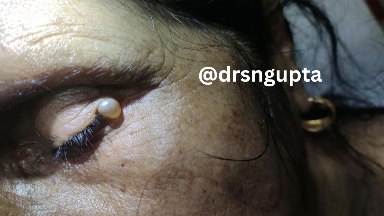
Hydrocystoma: Causes, Symptoms, Types, and Advanced Treatment Options
Hydrocystoma (also spelled hidrocystoma) is a rare, benign (non-cancerous) cystic lesion that originates from the sweat glands.Most commonly appearing on the face—particularly on or near the eyelids, cheeks, and forehead—these small, fluid-filled dome-shaped bumps can be a significant cosmetic concern.While hydrocystomas do not pose a direct threat to overall health or carry a risk of malignant transformation, their appearance can easily mimic serious skin conditions, including skin cancers.
Understanding hydrocystoma, its distinct subtypes, how it is diagnosed, and the available medical and surgical interventions can clarify what to expect during diagnosis and treatment.
What Is a Hydrocystoma?
A hydrocystoma is a benign, adnexal skin tumor formed by the retention of fluid within a blocked or dilated sweat gland duct.These cysts typically manifest as smooth, flesh-colored, translucent, or bluish nodules.They vary in size from a few millimeters to over a centimeter across.
Hydrocystomas are categorized into two primary histological classifications based on the type of sweat gland from which they originate:
- Eccrine Hydrocystoma:Originates from eccrine sweat glands, which are distributed across almost the entire human body and responsible for thermo-regulatory sweating.
- Apocrine Hydrocystoma:Originates from apocrine sweat glands, which are clustered in specific areas rich in hair follicles, such as the eyelids (specifically the glands of Moll), axillae, scalp, and groin.
Types of Hydrocystoma: Eccrine vs. Apocrine
Understanding the differences between eccrine and apocrine hydrocystomas is crucial for accurate clinical assessment and targeted therapy.
| Feature | Eccrine Hydrocystoma | Apocrine Hydrocystoma |
| Origin | Eccrine sweat gland duct | Apocrine sweat gland (e.g., Glands of Moll) |
| Etiology | Sweat duct retention/obstruction | Proliferation/adenomatous enlargement |
| Lesion Count | Single or multiple (Robinson type) | Usually solitary (Smith type) |
| Typical Size | Small (1 mm to 6 mm) | Larger (3 mm to 15 mm) |
| Color | Clear, flesh-colored, pale blue, amber | Dark blue, purple, reddish, or skin-colored |
| Primary Location | Periorbital region, cheeks, forehead | Eyelid margin, inner canthus, head, neck |
| Environmental Response | Size increases with heat and humidity | Unaffected by external heat/sweating |
1. Eccrine Hydrocystoma
Eccrine hydrocystomas result from the retention of sweat caused by an obstructed sweat duct. When the body produces sweat, the fluid becomes trapped, causing the duct to dilate into a fluid-filled cyst.
- Seasonal Variance: Eccrine hydrocystomas often display seasonal behavior. They tend to expand or become more visible in warm, humid weather or during physical exertion, and may diminish or temporarily vanish in colder environments.
- Clinical Variants: They can appear as single lesions or as multiple papules spread across the face—a presentation known as the Robinson-type eccrine hydrocystoma.
2. Apocrine Hydrocystoma
Apocrine hydrocystomas are hyperplastic outgrowth or true benign tumors (adenomas) rather than simple retention cysts.
- Appearance:These cysts are usually solitary, larger, and firmer than eccrine cysts.They frequently present with a dark bluish or dark brown hue.This deep pigmentation is caused by lipofuscin granules in the fluid and the Tyndall phenomenon—the scattering of light through the cystic fluid.
- Location:They show a strong preference for the eyelids, specifically near the inner canthus along the eyelashes. Unlike eccrine hydrocystomas, apocrine lesions do not fluctuate in size based on temperature changes.
Causes and Risk Factors
The precise cellular triggers leading to hydrocystoma development remain a topic of medical research, but several mechanisms and contributing factors have been identified:
- Ductal Blockage:Mechanical obstruction of the sweat duct due to localized inflammation, trauma, heavy cosmetics, or skin debris can prevent normal sweat drainage.
- Environmental Heat and Humidity: High ambient temperatures trigger increased sweat production. In individuals prone to ductal blockage, hyperhidrosis (excessive sweating) worsens fluid accumulation within the cyst wall.
- Gender and Age:Hydrocystomas are most frequently diagnosed in adults aged between 30 and 70. Multiple eccrine hydrocystomas are significantly more common in women, whereas solitary lesions show an equal distribution between sexes.
- Genetic and Syndromic Associations: While the majority of hydrocystomas occur spontaneously as isolated lesions, multiple apocrine or eccrine hydrocystomas can occasionally be linked to rare genetic conditions, such as Schöpf-Schulz-Passarge syndrome (a rare ectodermal dysplasia) or Goltz-Gorlin syndrome.
Common Symptoms and Visual Characteristics
Hydrocystomas are almost always asymptomatic from a physiological standpoint. They do not typically cause pain, itching, or inflammation unless secondary infection occurs or their physical presence mechanically interferes with nearby structures.
Common clinical features include:
- Smooth, Dome-Shaped Bumps:Soft to firm, fluctuant papules or nodules on the skin surface.
- Translucency: Cysts often exhibit a glassy, clear, or bluish appearance due to contained fluid.
- Location Sensitivity:When situated along the eyelid margin (glands of Moll), larger cysts can cause visual field obstruction, a sensation of heavy eyelids, or mild ocular irritation.
- Slow Growth:These lesions typically grow very slowly over years and persist indefinitely without treatment.
How Is a Hydrocystoma Diagnosed?
Because hydrocystomas can resemble other dermatological lesions, accurate medical evaluation is necessary.
+-------------------------------------------------------------------+
|                     CLINICAL PRESENTATION                         |
|         Asymptomatic translucent/bluish facial papule             |
+-------------------------------------------------------------------+
                                  |
                                  v
+-------------------------------------------------------------------+
|                        DERMOSCOPY EVALUATION                      |
|       Non-invasive imaging assessing vessel structure & hue        |
+-------------------------------------------------------------------+
                                  |
                +-----------------+-----------------+
                |                                   |
                v                                   v
+-------------------------------+   +-------------------------------+
|  Clear Diagnostic Indicators  |   |   Atypical or Suspicious      |
|  (Homogenous halo/pseudocyst) |   |   (Nodular/vascular features) |
+-------------------------------+   +-------------------------------+
                |                                   |
                v                                   v
+-------------------------------+   +-------------------------------+
|   Targeted Treatment /        |   |   EXCISIONAL SKIN BIOPSY      |
|   Conservative Management     |   |   Histopathological Analysis  |
+-------------------------------+   +-------------------------------+
1. Dermoscopy
Dermatologists often use dermoscopy (a polarized magnifying light tool) as a non-invasive preliminary evaluation. Under dermoscopy:
- Eccrine hydrocystomas present as clear, vessel-free, homogenous pseudocysts surrounded by a pale halo.
- Apocrine hydrocystomas display a blue, pink, or yellow homogeneous area with smooth, arborizing vessels running across the outer dome.
2. Histopathology (Biopsy)
A definitive diagnosis requires a skin biopsy or excisional tissue evaluation. Microscopic examination clearly differentiates the two types:
- Eccrine Hydrocystoma:Shows a unilocular (single-cavity) dermal cyst lined by one or two layers of flattened cuboidal epithelial cells, lacking myoepithelial modifications.
- Apocrine Hydrocystoma:Displays a multilocular cyst with an inner layer of tall columnar secretory cells showing characteristic decapitation secretion (apocrine cellular shedding) and an outer layer of myoepithelial cells.
3. Differential Diagnosis
It is essential to rule out other skin conditions that present with similar facial bumps:
- Basal Cell Carcinoma (BCC): Specifically nodular or cystic BCC, which carries malignant potential and requires formal surgical clearance.
- Blue Nevi and Melanoma: Pigmented skin lesions that can mimic the dark blue color of apocrine hydrocystomas.
- Syringoma: Small, firm sweat duct tumors, usually skin-colored or yellowish.
- Milia & Epidermal Cysts:Keratin-filled cysts rather than fluid-filled sweat structures.
Treatment Options for Hydrocystoma
Because hydrocystomas are benign, medical intervention is not always strictly necessary.Treatment is usually sought for cosmetic reasons, recurrent inflammation, or mechanical impairment of vision.
Selection of treatment depends on whether the patient presents with a single cyst or multiple lesions.
1. Surgical Excision (Gold Standard for Solitary Lesions)
For a single apocrine or eccrine hydrocystoma, complete surgical excision with intact cyst wall removal is the most effective treatment.
- Procedure: Performed under local anesthesia, the surgeon carefully dissects the cyst intact away from surrounding delicate eyelid or facial tissue.
- Recurrence: If the cyst wall is completely excised intact, recurrence rates are extremely low.
2. Minimally Invasive Procedures
When surgical excision carries scar risk in high-visibility facial zones, alternatives exist:
- Puncture and Simple Drainage: Piercing the cyst with a sterile needle allows immediate fluid release. However, because the secretory lining remains intact, fluid almost always reaccumulates, leading to high recurrence rates.
- Electrodessication and Curettage: After puncturing the cyst, electric current is applied to cauterize the inner epithelial lining, preventing future fluid secretion.
- Laser Therapy: Carbon Dioxide ($\text{CO}_2$) lasers or Pulsed Dye Lasers (PDL) can vaporize or coagulate the cyst lining with minimal scarring.
- Chemical Sclerotherapy:Injection of sclerosing agents like hypertonic glucose or trichloroacetic acid (TCA) after cyst aspiration damages the secretory lining, inducing collapse and closure.
3. Medical and Topical Treatments (Ideal for Multiple Lesions)
Surgical removal of dozens of tiny cysts (multiple eccrine hydrocystomas) can cause unacceptable facial scarring. Medical management is preferred in these cases:
- Topical Anticholinergic Agents: Medications such as atropine cream, scopolamine, or glycopyrrolate suppress eccrine gland sweat production, leading to shrinkage of the cysts.
- Botulinum Toxin (Botox) Injections: Perilesional intradermal injections of Botulinum Toxin Type A block the release of acetylcholine at the neuromuscular and neuroglandular junctions. This stops sweat production in the treated zone, causing dramatic flattening of eccrine hydrocystomas that lasts several months.
- Topical Botulinum-like Peptides: Newer topical peptide formulations offer a non-invasive alternative to reduce sweat stimulation without injections.
Managing Hydrocystoma: Lifestyle and Prevention Tips
While genetic factors and sweat gland architecture cannot be modified, specific daily management strategies can help reduce flare-ups, particularly for individuals with eccrine hydrocystomas:
- Minimize Excessive Heat Exposure: Avoid prolonged exposure to saunas, hot showers, and direct heat sources that trigger intense facial sweating.
- Choose Climate-Controlled Environments: Maintain cool indoor temperatures during hot, humid summer months to lower ambient sweating triggers.
- Avoid Occlusive Cosmetics:Heavy, oil-based foundations or facial products can block sweat pores. Opt for light, non-comedogenic cosmetics.
- Resist Squeezing or Popping Cysts: Attempting to rupture a hydrocystoma at home introduces bacteria, leading to cellulitis, scarring, or localized tissue damage near the eyes.
Frequently Asked Questions (FAQs)
Is a hydrocystoma cancerous?
No, hydrocystomas are entirely benign (non-cancerous) growths of the sweat glands.They do not turn into skin cancer, though their appearance can resemble certain forms of skin malignancies.
Will a hydrocystoma go away on its own?
Generally, no. While eccrine hydrocystomas may shrink during cold winter months, the cyst wall remains and typically refills during warmer weather. Complete resolution usually requires targeted medical or surgical intervention.
Are hydrocystomas contagious?
No, hydrocystomas are non-infectious structural lesions caused by blocked or proliferating sweat glands. They cannot spread from person to person.
Does surgical removal leave a scar?
Any surgical incision can leave a fine scar; however, dermatologists and oculoplastic surgeons use precise micro-surgical techniques to minimize scar visibility, particularly around delicate areas like the eyelids.

Dr. S. N. Gupta is a renowned skin specialist and dermatologist in Prayagraj, with extensive experience in diagnosing and treating various skin, hair, nail and Sexually Transmitted Diseases conditions for last 28 years. Address Shree Balaji Medical Traffic Chauraha, Rajapur Prayagraj Mob 9415613743