
Book Appointment Now
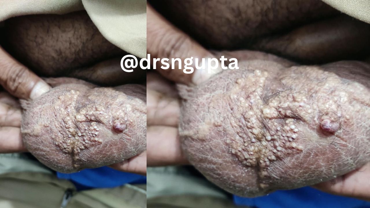
Lymphangioma- causes , symtoms and treatment
A lymphangioma—medically classified as a lymphatic malformation (LM)—is a rare, benign (non-cancerous) vascular anomaly of the lymphatic system. Occurring in approximately 1 in 4,000 live births, these growths form when embryonic lymphatic fluid channels fail to connect properly to the main venous network during fetal development. This leads to local fluid pooling, vessel enlargement, and cyst formation.
Though technically present at birth, lymphangiomas may remain undetectable until early childhood, with over 90% diagnosed before age two.
Causes and Pathophysiology
The underlying driver of congenital lymphangiomas is an isolated somatic mutation occurring during embryogenesis—meaning it is non-heritable and cannot be passed down through generations. Modern molecular genetics points to somatic gain-of-function mutations in the PIK3CA gene across a high percentage of lymphatic malformations. This gene regulates cell growth and vascular development; its hyperactivation leads to abnormal proliferation of lymphatic endothelial cells.
While most cases appear sporadically, lymphangiomas occasionally occur alongside underlying chromosomal syndromes, including:
- Turner Syndrome
- Noonan Syndrome
- Down Syndrome (Trisomy 21)
Acquired Lymphangiomas: Less commonly, adults develop secondary lymphatic malformations (often called lymphangiectasia). These stem from physical disruption or obstruction of previously normal lymphatic channels—frequently secondary to surgical lymph node dissection, pelvic radiation therapy, severe tissue trauma, or chronic infections.
Major Types and Classifications
Modern medical consensus categorizes lymphangiomas into three primary subtypes based on cyst vessel size, depth, and histological characteristics.
| Type | Cyst Size | Common Locations | Clinical Features |
| Macrocystic(Cystic Hygroma) | Large cysts (> 2 cm) | Posterior neck, axilla (armpit), chest wall | Soft, smooth, transilluminating masses under normal or bluish skin. Responds well to sclerotherapy. |
| Microcystic(Lymphangioma Circumscriptum) | Small cysts (< 2 cm) | Tongue, oral mucosa, skin of trunk and limbs | Cluster of translucent or dark red vesicles resembling “frog spawn”. Highly infiltrative tissue pathways. |
| Mixed | Variable (combines macro & micro) | Head, neck, cheek, and deep visceral spaces | Shows both discrete fluid-filled pockets and surrounding diffuse, infiltrated tissue. |
Symptoms and Clinical Presentation
Symptoms vary depending on the lesion’s size, depth, and proximity to vital organ structures.
- Superficial Skin Manifestations: Small, fluid-filled skin bumps ranging from translucent to pink, dark red, or purple (if tiny blood vessels leak into the cyst).
- Deep Soft-Tissue Masses: A painless, spongy, compressible lump beneath the skin that slowly enlarges alongside the child’s natural growth.
- Airway or Swallowing Impairment: Cervical (neck) or mediastinal (chest) lesions can compress the trachea or esophagus, triggering stridor, dyspnea, or dysphagia.
- Rapid Expansion: Viral or bacterial upper respiratory infections can cause a sudden, painful swelling of the mass due to fluid buildup or internal bleeding.
Diagnostic Procedures
An accurate diagnosis distinguishes lymphatic malformations from hemangiomas, venous malformations, and soft-tissue sarcomas.
- Prenatal Ultrasound: High-resolution routine fetal ultrasounds can detect large macrocystic lesions (cystic hygromas) as early as the late first or second trimester.
- Postnatal Physical Exam: Evaluation using transillumination—shining a light source through the soft tissue mass—to verify whether the fluid composition is clear lymph.
- Magnetic Resonance Imaging (MRI): The gold standard imaging modality. MRI pinpoints exact anatomical borders, depth, tissue infiltration, and involvement of adjacent neurovascular bundles without ionizing radiation.
- Ultrasound & CT Scans: High-frequency Doppler ultrasound confirms slow-flow fluid dynamics without arterial or high-pressure venous activity.
Modern Treatment Options
Treatment selection depends on lesion subtype (macrocystic vs. microcystic), location, size, and symptom severity.
1. Active Surveillance (“Watch and Wait”)
Asymptomatic, small lesions that present no functional risk or aesthetic concern are monitored through regular clinical imaging examinations.
2. Image-Guided Sclerotherapy
Sclerotherapy is the primary non-surgical intervention for macrocystic malformations. An interventional radiologist drains the cyst under ultrasound guidance and injects a sclerosant agent (such as Bleomycin, OK-432, Doxycycline, or Polidocanol). This irritates the endothelial lining, causing the cyst walls to collapse, scar, and permanently shrink.
3. Targeted Medical Therapy
For extensive, deep, or microcystic malformations where surgery carries high nerve damage risks, oral Sirolimus (Rapamycin)—an mTOR inhibitor—has emerged as an effective targeted therapy. It inhibits lymphatic vessel endothelial cell growth, significantly reducing lesion volume and symptoms.
4. Surgical Excision
Complete surgical resection is often reserved for localized microcystic lesions or cases where sclerotherapy fails. Because these malformations lack defined anatomical capsules and weave around surrounding nerves and muscle tissue, complete removal is crucial to prevent recurrence.
Complications
Overall long-term survival for isolated lymphangiomas is excellent, but management requires awareness of potential complications:
- Infection (Cellulitis): Intracystic bacteria can cause localized warmth, pain, fever, and sepsis if untreated.
- Intracystic Hemorrhage: Minor trauma can cause fragile blood capillaries within the cyst walls to rupture, leading to sudden, painful enlargement.
- Recurrence Rates: If microcystic tissue components are incompletely excised, local recurrence rates range between 15% and 40%.

Dr. S. N. Gupta is a renowned skin specialist and dermatologist in Prayagraj, with extensive experience in diagnosing and treating various skin, hair, nail and Sexually Transmitted Diseases conditions for last 28 years. Address Shree Balaji Medical Traffic Chauraha, Rajapur Prayagraj Mob 9415613743